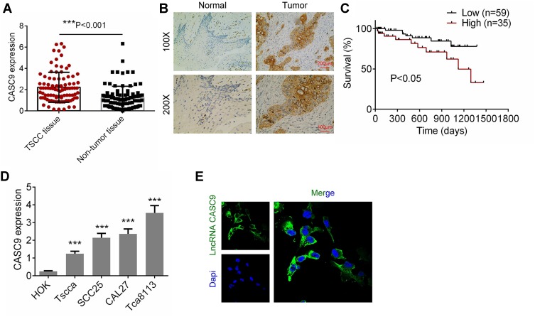
Figure 1.
The expression of CASC9 in TSCC tissues and cell lines. (A) The mRNA expression of CASC9 in 41 paired TSCC tissues and matched adjacent non-TSCC tissues was detected by RT-qPCR. (B) The expression of CASC9 in TSCC tissues and matched adjacent non-TSCC tissues was detected by ISH. (C) Survival rates of patients with TSCC with high and low CASC9 by Kaplan-Meier survival analysis. (D) The expression of CASC9 in TSCC cell lines (CAL27, Tca8113, Tscca and SCC25) and human oral keratinocytes (HOK) were determined by RT-qPCR. (E) The cellular sublocalization of CASC9 was evaluated by immunofluorescence assay. Data is from three independent experiments and expressed as mean ± SD. ***P<0.001.

