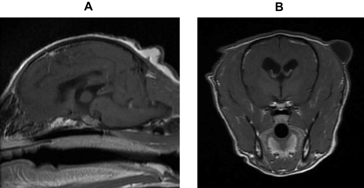
Figure 7.
A case of a dog with parapituitary meningioma. A twelve-year-old, castrated Toy Poodle was referred with complaints of polyuria, polydipsia, polyphagia, abdominal enlargement, dorsal alopecia, and poor wound healing. The dog was diagnosed with Cushing’s syndrome and diabetes mellitus based on the results of the endocrinological tests and abdominal ultrasound. Upon MRI examination, PBR was 0.27, and MRI-grading was II. TSS was performed and a diagnosis of meningioma was made. (A) Gd-T1 weighted sagittal image. (B) Gd-T1 weighted transverse image.

