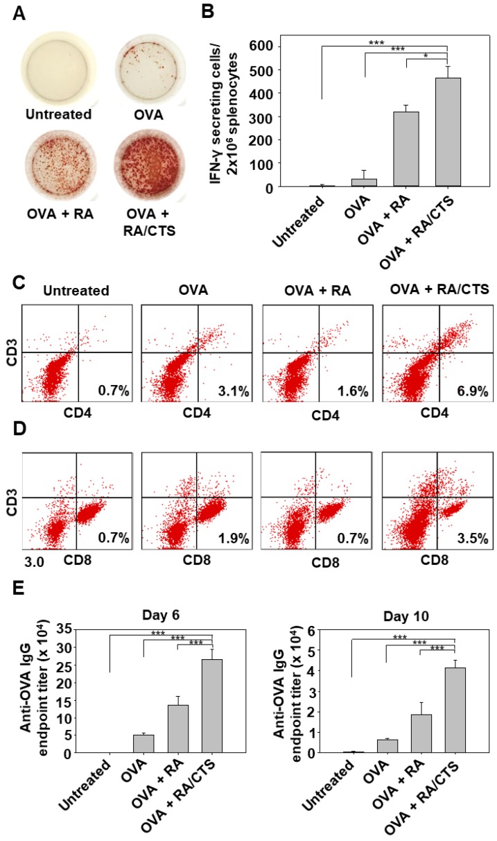
Figure 7.
Post-vaccination humoral and cellular immune responses. Naïve mice were vaccinated three times with OVA in various formulations at 4-day intervals. Five days after the last vaccination, mice were inoculated with B16-OVA tumor cells (3 × 10⁵). Splenocytes were isolated 2 days after tumor challenge for ex vivo IFN-γ ELISPOT (A). IFN-γ–secreting cells were counted using a stereomicroscope (n = 4); * p < 0.05; *** p < 0.005) (B). Tumor-infiltrating T cells in tumors were analyzed for CD3(+)CD4(+) T helper cells (C) and CD3(+)CD8(+) cytotoxic T cells (D) by flow cytometry on day 27. Blood samples were collected 6 and 10 days after inoculation for serum anti-OVA IgG titration. Anti-OVA IgG levels were measured by ELISA in serum samples diluted at different ratios (n = 5) (*** p < 0.005) (E).

