Figure 2.

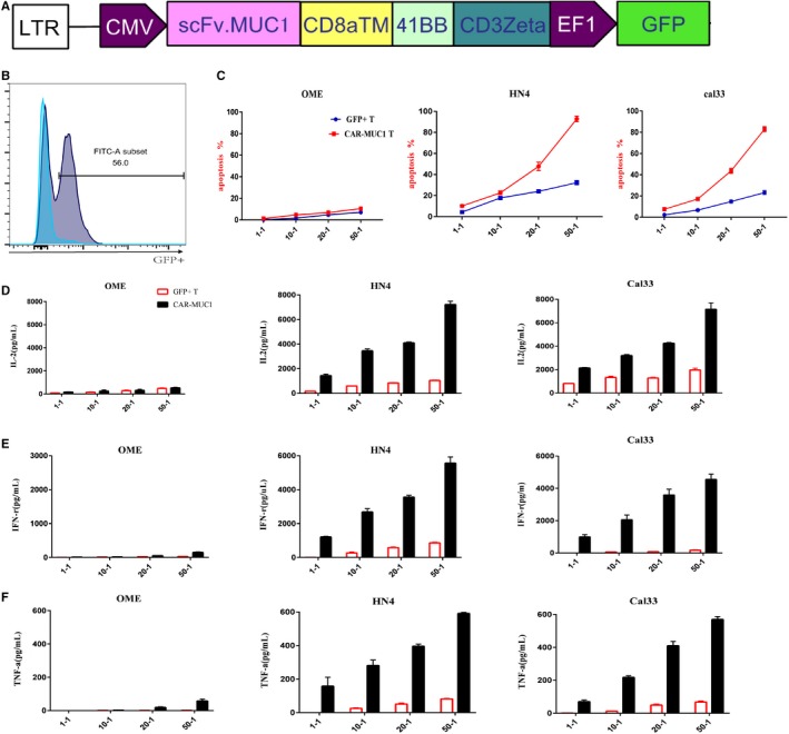
Chimeric antigen receptor‐mucin 1(CAR‐MUC1) T cells specifically killed MUC1+ HNSCC cell lines in vitro. A, Construction of CAR‐MUC1. B, Transfect efficiency of CAR‐T cells. Flow cytometry tested the positive rate of GFP compared with nontransfection. (The blue is control group and gray is positive group.) C, Flow cytometry tested the lysis of different tumor cells by CAR‐MUC1 T cells and GFP+ T cells, respectively. CAR‐MUC1 T and GFP+ T cells were coculture with tumors cell from OME, HN4, Cal33 for 6 h, respectively. The results were the sum of Annexin5 single‐positive rate (early apoptosis) and PI, Annexin5 double‐positive rate (late apoptosis). D‐F, IL‐2, IFN‐γ, and TNF‐α secretion of CAR‐MUC1 T cells and GFP+ T cells in coculture supernatants after different E/T ratio were measured by ELISA assay. Each trial was repeated three times, the T cells came from three different healthy donors. Both CAR‐T and GFP+ T cells in each trial came from the same volunteer
