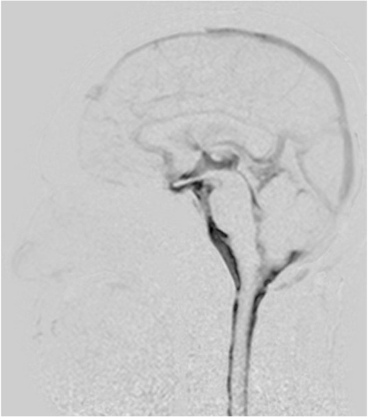
Fig. 1.

Mid-sagittal image of cerebrospinal fluid (CSF) motion in a 48-year-old healthy male volunteer visualized using dynamic improved motion sensitized driven-equilibrium steady-state free precession (Dynamic iMSDE SSFP). Generally, the dark area on the grayscale images shows vigorous turbulent CSF motion. The dark image contrast is achieved by signal attenuation induced by irregular or turbulent CSF motions in each site compared with the surrounding areas where CSF moves relatively mildly in the Dynamic iMSDE SSFP image. In this healthy individual, increased signal intensity is shown in the area of the chiasmatic cistern, anterior part of the brainstem, cisterna magna, Sylvian aqueduct, and third ventricle. Note increased signal intensity in the anterior horn. This may indicate vigorous CSF motion that is transmitted from the third ventricle to the anterior horn through the foramen of Monro.
