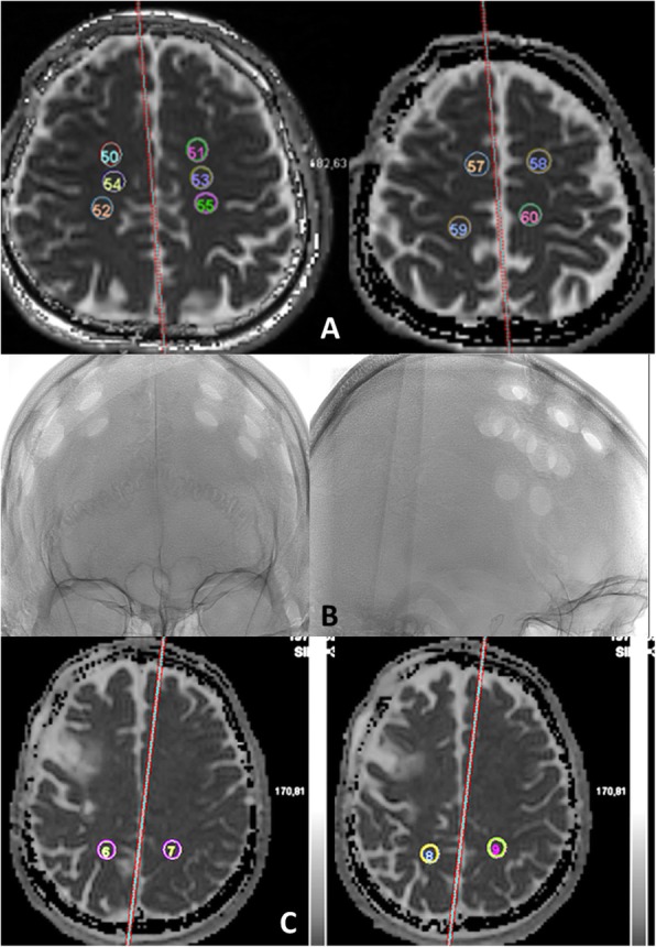
Fig. 1.

Template of the location of regions of interest (ROI) in normal-appearing white matter (Anterior ROIs (a); posterior ROIs (c) with an example of burr-holes location on angiography (b) to show that posterior ROIs were far from burr holes). ADC sequences passing through frontal white matter at the level of centrum semiovale. A minimum of 4 ROIs were placed in each hemisphere, but if possible, 1 or 2 more ROIs were added. The template was copied after surgery
