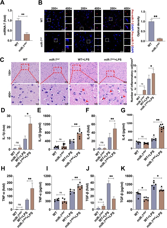
Fig. 2.
MiR-7 deficiency aggravates the pathology of BTI model. Animal experiments were performed according to Fig. 1a. a The expression level of miR-7 in brain tissue was determined by Real-time PCR assay and calculated, respectively. b The expression level of pre-miR-7-2 was determined by FISH (n = 4 per group). WT mice and miR-7def mice were intraperitoneally injected with LPS and control group with PBS. After 12 h, c the pathology of brain tissue was observed by H&E staining (n = 3 per group). Arrows in c indicate hemorrhage foci. d–k The expression levels of IL-1β, IL-6, TNF-α, and TGF-β were analyzed by real-time PCR and ELISA assay (n = 6 per group; one-way ANOVA, *P < 0.05, **P < 0.01)

