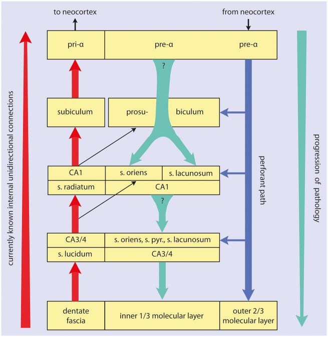
FIGURE 1.
Summary diagram of temporal lobe allocortical connectivities. The direction of the main efferent projections is indicated by the red arrow at the far left. Granule cells of the dentate fascia project via mossy fibers to CA3 and CA4 projection neurons. Axons of these cells contribute to the alveus and give off Schaffer collaterals to CA1 pyramidal cells. These, in turn, project via the ammonic-subicular pathway to the prosubiculum/subiculum, and subicular efferents reach the deep layer pri-α of the entorhinal region (short red arrows). Outer layers of the entorhinal and transentorhinal regions project via the perforant path (blue arrow) to the Ammon’s horn and predominantly to the dentate fascia. The progression of the sAD-associated pathology is indicated by the green arrow at the far right. Shorter green arrows between the boxes indicate the here hypothesized projections from pre-α cells to the prosubiculum and both the stratum oriens and stratum lacunosum of CA1, as well as the hypothetical projections from prosubicular and/or CA1 pyramidal cells to the stratum oriens, stratum lacunosum, and to thorny excrescences of CA3/CA4 projection neurons. Question marks indicate uncertainties regarding the existence of these connections. A known pathway is given off from CA3/CA4 mossy cells to the inner one-third of the dentate molecular layer.

