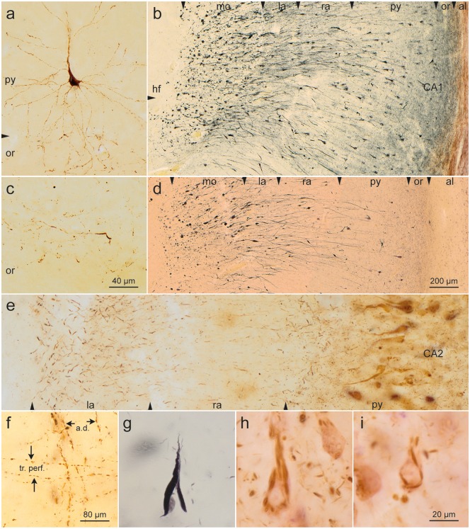
FIGURE 4.
Somatic alterations of CA1 projection neurons. (a, c) Early development of abnormal tau in a CA1 pyramidal cell. Note changes in distal segments of the basal dendrites that reach into the stratum oriens. (c) A portion of the stratum oriens in an early phase of development (AT8, case 23, male, age 64, NFT II, Aβ 0). (b, d) CA1 pyramidal cells are prone to develop alterations of their apical dendrite. Their terminal portions in the molecular layer (mo) develop, extensions interconnected by thread-like segments (AT8, blue chromogen). The same case seen in Gallyas silver-iodide (case 7, male, age 69, NFT I, Aβ 0). In both micrographs, the lateral ventricle (not shown) would be located at the far right (see also Fig. 3a). (e) Apical dendrites of CA2 pyramidal cells show a less conspicuous but basically similar change with thickenings of their terminal segments (AT8, case 26, female, age 82, NFT II, Aβ 1). (f) Stratum lacunosum of CA1. Apical dendrites of CA1-pyramidal cells (a. d.) and involved axons of the perforant path (tr. perf.) (AT8, case 22, male, age 64, NFT II, Aβ 0). Altered CA1 cells. (g) Mature argyrophilic neurofibrillary tangle of a CA1 pyramidal cell (Gallyas silver-iodide staining). (h, i) Flake-like formation of fibrillized tangle in CA1 pyramidal cells. Note the absence of perinuclear pretangle material (AT8, case 60, female, age 84, NFT IV, Aβ 2). Scale bars: (c) = 40 μm, also applies to (a); (d) = 200 μm, also applies to (b); (f) = 80 μm; (i) = 20 μm, also applies to (e), (g), and (h).

