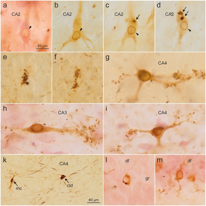
FIGURE 5.
Pretangles in CA2 cells developed gradually and were evenly distributed throughout the somatodendritic compartment, consistently showing perinuclear tau accumulation (arrowheads). (c, d) A few rounded accumulations of densely packed abnormal tau (arrows) occasionally occurred in the cytoplasm [(a) case 60, female, age 84, NFT IV, Aβ 2; (b–d) case 75, male, age 80, NFT V, Aβ 2]. (e, f) The earliest changes in CA3/CA4 were small AT8-immunoreactive dots that occur in isolation and resemble dendritic excrescences (case 75, male, age 80, NFT V, Aβ 2). These cells showed characteristic excrescences along their dendrites and, similar to CA2 cells, exhibited a zone of perinuclear tau accumulation [(g) case 45, female, age 83, NFT III, Aβ 0; (h, i) case 60, female, age 84, NFT IV, Aβ 2]. (k) The first cells involved in CA4 usually belonged to the type of multipolar pigment-laden cells with long and smoothly contoured dendrites without excrescences (arrow: cld). For comparison, a typical CA4 mossy cell is shown at the left (arrow: mc) (case 45, female, age 83, NFT III, Aβ 0). The last cells to develop changes were the granule cells (gr) of the dentate fascia (df). These cells showed a uniformly distributed AT8-positive tau in the cytoplasm as well as perinuclear tau accumulations. (l) Rarely, AT8-positive dots were seen in the cytoplasm. (m) Distal dendritic segments did not show spindle-shaped enlargements (case 60, female, age 84, NFT IV, Aβ 2). Scale bars: (a–i, l, m) = 20 μm; (k) = 40 μm.

