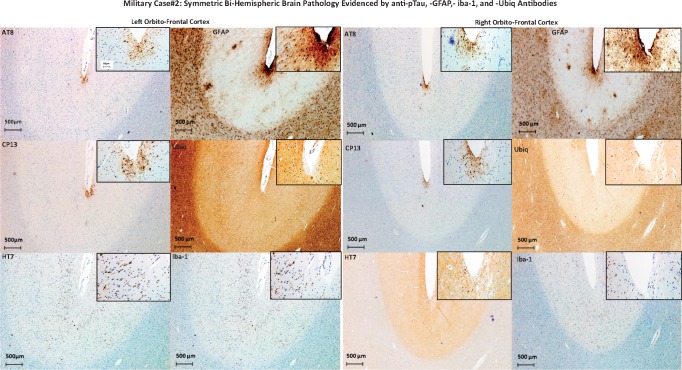
FIGURE 3.
Military Case 2: Symmetric bihemispheric brain pathology evidenced by anti-pTau, -GFAP, -Iba-1, and -Ubiq antibodies. The figure shows left and right orbito-frontal cortical areas of Case 2 brain stained for pTau (AT8, CP13), all Tau (HT7), astroglial cells (GFAP), ubiquitin (Ubiq), and microglia (Iba-1). Each image contains a larger image at lower magnification and an inset (upper right corner of each image) of the same cortical area at higher magnification.

