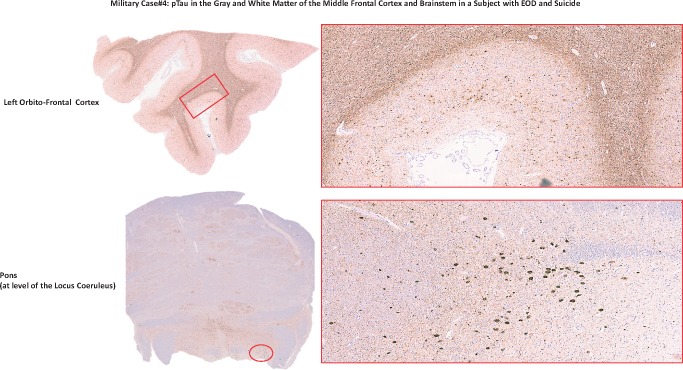
FIGURE 5.
pTau in the gray and white matter of the middle frontal cortex and brainstem in a subject with EOD and suicide. The figure shows left orbito-frontal cortex and pons area of the Case 4 brain stained for pTau (AT8). Photographs were taken at lower (left) and higher (right) magnification. The red rectangle and circle on the lower magnification photographs correspond to the area on the right photographed at higher magnification.

