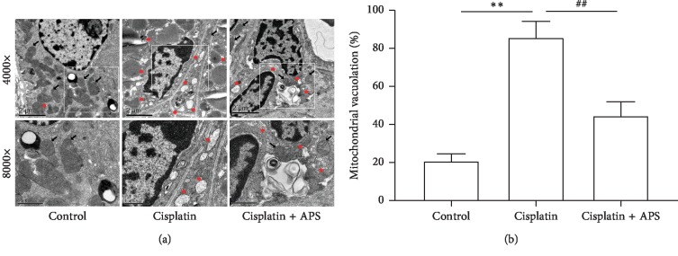
Figure 5.
APS ameliorates the cisplatin-triggered mitochondrial damage in mice. (a) Transmission electron microscopy images of the mitochondrial morphology changes of mice renal (black arrow indicates normal mitochondria; red asterisk indicates damaged mitochondria; bar = 2 μm and bar = 1 μm). (b) Quantitative analysis of mitochondrial vacuolation. Data are means ± SEM (n = 3). ∗∗P < 0.01 versus the control group; ##P < 0.01 versus the cisplatin alone-treated group.

