Figure 5.

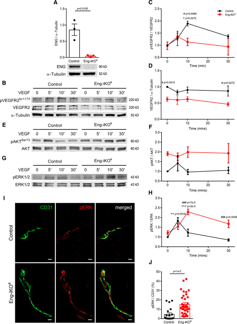
Endothelial cells (ECs) show altered VEGF (vascular endothelial growth factor) signaling responses following loss of ENG (endoglin). A, ECs cultured from Rosa26-CreERT2;Engfl/fl mice were transiently exposed to 4OH-tamoxifen to deplete ENG protein, which is measured by Western blot. Data were analyzed by Student unpaired t test (n=3). B–D, Cells exposed to VEGF (10 ng/mL) for 0, 5, 10, and 30 min and assessed by Western blot show a reduced VEGFR2 (VEGF receptor 2) phosphorylation response and reduced overall VEGFR2 levels compared with control ECs. E and F, Basal pAKT (phospho-protein kinase B) activity appears enhanced in Eng-iKO ECs compared with controls. G and H, Eng-iKO ECs have an increased and prolonged pERK (phospho-extracellular signal-regulated kinase) response compared with control cells. Molecular weights of named proteins are predicted based on molecular weight standards. Data in C, D, F, and H were analyzed using 2-way ANOVA with Bonferroni correction. Asterisks (*) indicate significant changes with respect to baseline for each genotype, while hashes (#) indicate significant differences between control and Eng-iKO ECs; the full set of P is summarized in Online Table VI (n=3–5/group). I, Increased levels of pERK in ECs of pubic symphysis of Eng-iKOe compared with control mice (scale bar=10 µm). J, Quantification of pERK vascular coverage from individual vessels from control (6 mice, n=19 vessels) and Eng-iKOe (4 mice, n=35 vessels) mice. Data analyzed by Mann-Whitney U test. AKT indicates protein kinase B; CD31, cluster of differentiation 31; and ERK, extracellular signal-regulated kinase.
