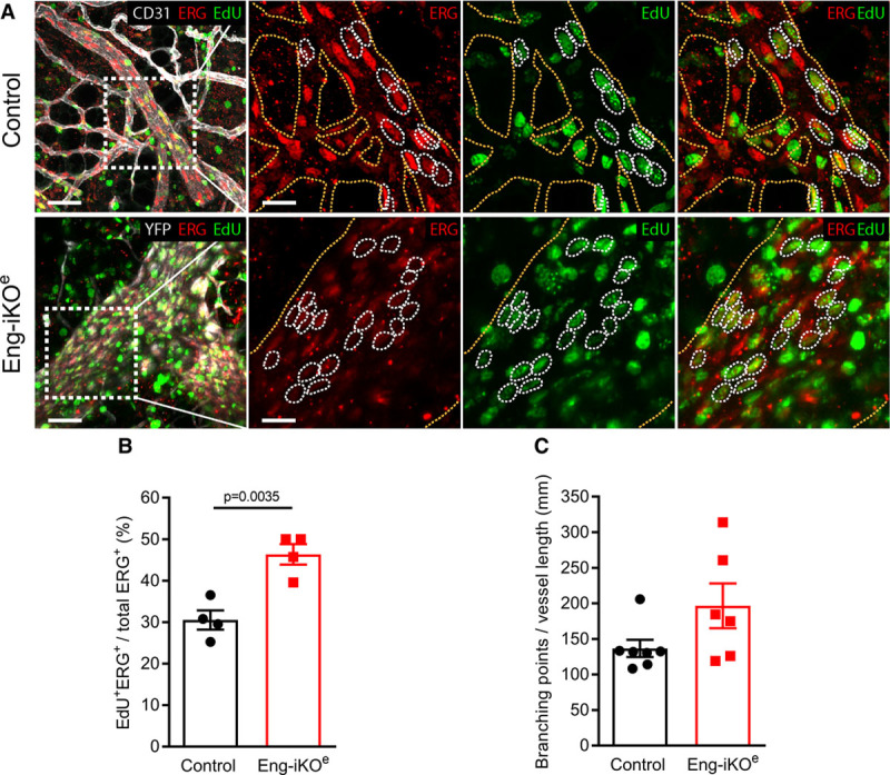
Figure 6.

Endothelial cell (EC) proliferation of the adult vasculature is promoted by endoglin deletion. A, Vessels of the pubic symphysis of control (top) and Eng-iKOe (bottom) mice, exposed to repeated 5-ethynyl-2-deoxyuridine (EdU) injections over 2 wk, stained for CD31 (cluster of differentiation 31) or YFP (yellow fluorescent protein; ECs, white), ERG (erythroblast transformation-specific transcription factor; EC nuclei, red), and EdU (proliferating cells, green). Left, Merged overviews. Scale bars=50 µm. Right, Boxed areas in left. Scale bars=20 µm. Yellow dotted lines outline the vasculature, and white dotted lines indicate proliferating ECs (ERG+ and EdU+, double positive). B, Quantification of accumulated EC proliferation over 2 wk demonstrates an increase from 31% proliferating ECs in control (n=4 mice) to 45% in Eng-iKOe (n=4 mice). Data were analyzed by Student unpaired t test. C, Quantification of average EC size calculated from the number of ERG+ cells per vessel shows a trend toward an increased EC size in Eng-iKOe mice (n=6) compared with controls (n=7). Data were analyzed by Mann-Whitney U test.
