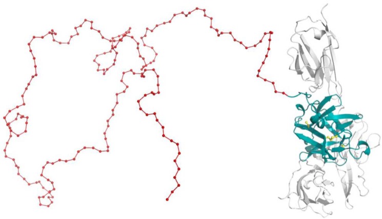
Figure 1.
Three-dimensional structure of PASylated IL-1Ra. Molecular model of IL-1Ra (teal) with an N-terminal PAS extension (here, 200 residues, red) in arbitrary random conformation. Association with the extracellular domain of IL-1R1 (gray) is indicated (based on the crystal structure of the complex, PDB code 1IRA (67)). The four Cys side chains of IL-1Ra are highlighted (yellow); two of them are engaged in a structural disulfide bridge.

