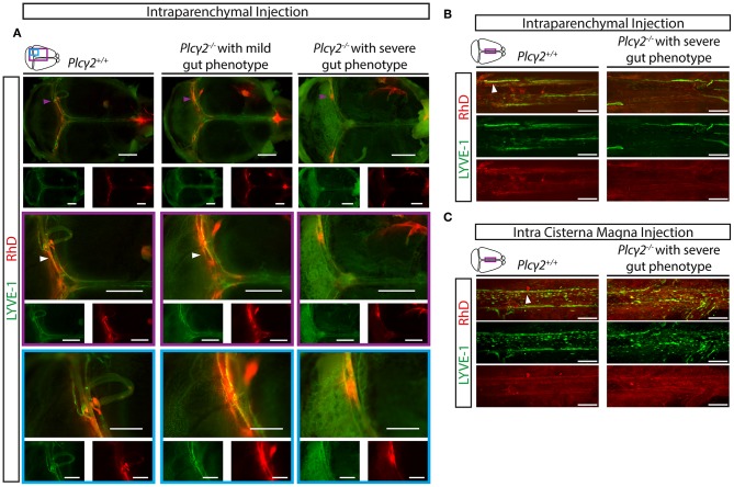
Figure 9.
Uptake and transport of labeled macromolecules by the meningeal lymphatics in Plcγ2+/+ and Plcγ2−/− mice. (A) Uptake and transport of 70 kDa RhD by the meningeal lymphatic vessels after intraparenchymal injection of young adult Plcγ2+/+, Plcγ2−/− mice displaying mild or severe gut phenotype. Representative images are shown of whole-mount anti-LYVE-1 immunostaining of meninges of young adult mice (n = 8 for Plcγ2+/+ mice; n = 3 for Plcγ2−/− mice showing mild gut phenotype, n = 5 for Plcγ2−/− mice displaying severe gut phenotype). Purple arrowheads point to spots with a high intensity for macromolecule uptake, white arrowheads show meningeal lymphatic vessels draining RhD. Bars, 1,000 μm. (B,C) Confocal images are shown of region adjacent to the superior sagittal sinus of whole-mount immunostained meninges of young adult Plcγ2+/+ and Plcγ2−/− mice after intraparenchymal (B) or intra cisterna magna (C) injection of 70 kDa RhD (intraparenchymal injection: n = 3 for Plcγ2+/+ mice; n = 3 for Plcγ2−/− mice displaying severe gut phenotype; intra cisterna magna injection: n = 3 for Plcγ2+/+ mice; n = 2 for Plcγ2−/− mice displaying severe gut phenotype). White arrowheads show meningeal lymphatic vessels draining RhD. Bars, 200 μm.

