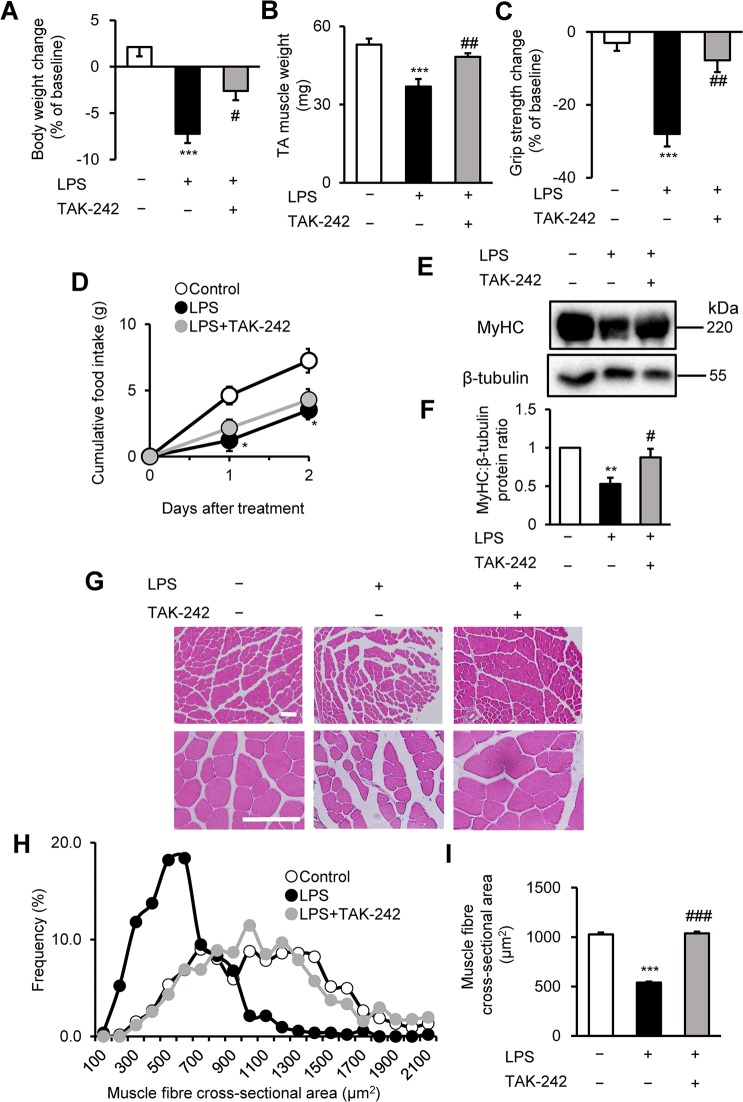
Figure 3.
TAK-242 reduces LPS-induced muscle atrophy and weakness in mice. (A–D) Wild-type C57BL/6 mice (8–12-week-old males) were injected with vehicle (PBS containing 0.9% DMSO) or TAK-242 (3 mg/kg) and then with PBS or LPS (1 mg/kg) 1 h later. After 2 days, mice were assessed for body weight (A), TA muscle weight (B), and grip strength (C). Food intake (D) was measured every 24 h up to 48 h. N = 4–5/group. (E,F) Western blot analysis (E) and quantification (F) of MyHC expression in TA muscles at 2 days after administration of LPS (1 mg/kg) and TAK-242 (3 mg/kg) as described for (A–D). Data were normalised to β-tubulin protein levels, and the ratio in vehicle control-treated mice was set at 1.0. N = 7–8/group. Full-length blots are presented in Supplementary Figure S6. (G–I) Representative images of H&E-stained TA muscle sections (G) and quantification of the distribution (H) and mean (I) cross-sectional areas of TA muscle fibres at 2 days after administration of LPS (1 mg/kg) and TAK-242 (3 mg/kg) as described for (A–D). The cross-sectional area of TA muscle fibres was measured as described in the Methods. Scale bar, 100 μM. N = 506–523/group. For all panels, data are presented as the mean ± s.e.m. ***p < 0.001, **p < 0.01, *p < 0.05 vs vehicle control, ###p < 0.001, ##p < 0.01, #p < 0.05 vs LPS-treated group by one-way ANOVA followed by Tukey’s honest significant difference test.

