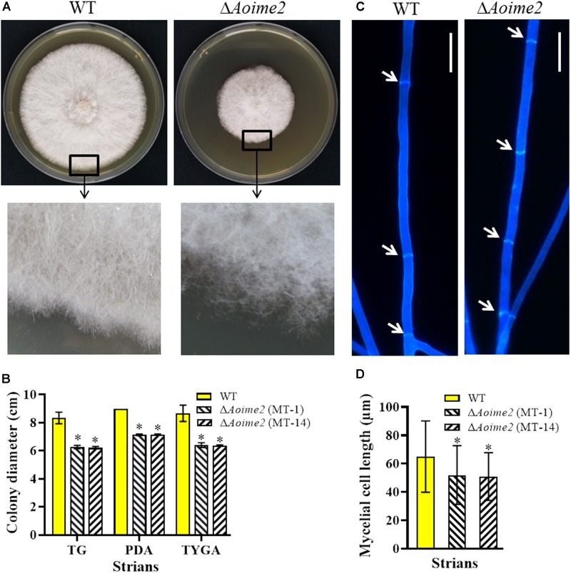
FIGURE 1.
Comparison of mycelial growth, morphology, and septum formation between wild type (WT) and ΔAoime2 mutants. (A) Colony morphology (upper panel) and hyphae (lower panel) of WT and ΔAoime2 mutant strains incubated on TYGA medium for 5 days at 28°C. (B) Colony diameters of WT and ΔAoime2 mutants incubated on PDA, TYGA, and TG media for 7 days. (C) Hyphal septa of WT and mutants were stained with 20 μg/mL calcofluor white (CFW) after the fungal strains were incubated on CMY medium for 7 days. Arrows: hyphal septa. Bar = 10 μm. (D) Comparison of mycelial cell size of WT and mutants; 60 mycelial cells were randomly selected, and the distance between two hyphal septa was measured using ImageJ software. Error bars: SD from 60 replicates; asterisk: significant difference between mutant and WT (Tukey’s HSD, p < 0.05).

