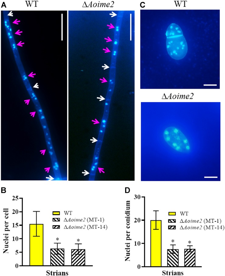
FIGURE 2.
Mycelial and conidial cell nuclei in WT versus ΔAoime2 mutant strains. (A) Hyphae of WT and ΔAoime2 mutant strains were stained with CFW and DAPI after the fungal strains were grown for 7 days on CMY medium; samples were examined using an inverted fluorescence microscope. White arrows: septa; pink arrows: cell nuclei, the strong stained and bright points are identified as nuclei. Bar = 20 μm. (B) Comparison of mycelial cell nuclei between WT and mutant strains; 30 mycelial cells were randomly selected and the nucleus in each cell was counted separately. (C) Conidia of WT and ΔAoime2 mutant strains were stained with CFW and DAPI. Bar = 10 μm. (D) Comparison of conidial cell nuclei between WT and mutant strains; 30 conidia were randomly selected for counting cell nuclei. Error bars: SD from 30 replicates; asterisk: significant difference between mutant and WT (Tukey’s HSD, p < 0.05).

