
FIGURE 4.
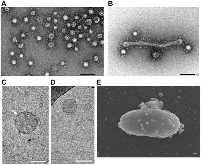
Morphological characterization of EMVs from S. vesiculosa HM13. (A) Negative-stained TEM image of EMVs from S. vesiculosa HM13 grown at 4°C. (B) Negative-stained TEM image of tube-like structure observed in the EMV fraction of S. vesiculosa HM13 grown at 4°C. (C,D) Cryo-EM images of EMVs from S. vesiculosa HM13 grown at 4°C. An arrow indicates EMV surrounded by a high electron density region. (E) FE-SEM image of the S. vesiculosa HM13 cell grown at 18°C showing many buds appearing on the cell surface. Bars represent 100 nm.