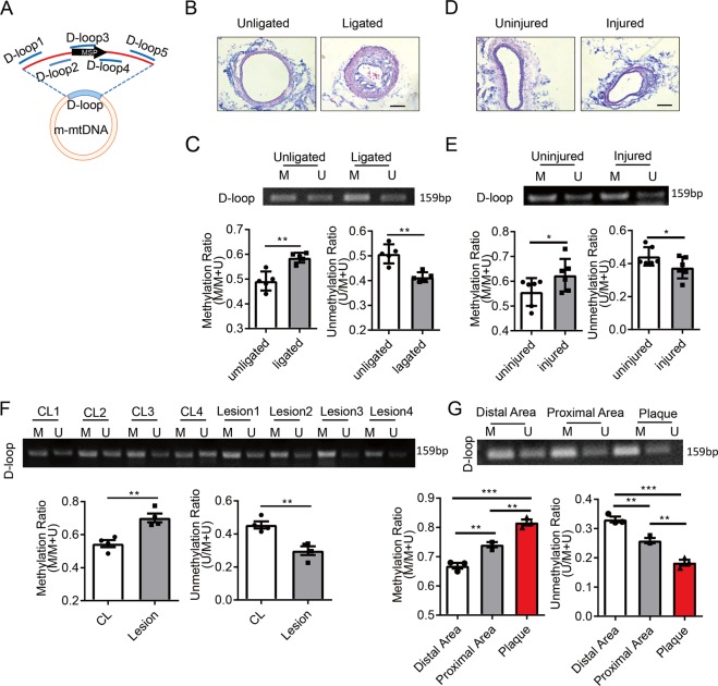
Fig. 5. Mitochondrial D-loop region is hypermethylated in smooth muscle cells from arterial stenotic and occlusive diseases.
a Schematic diagram of mouse mitochondrial DNA (m-mtDNA). Locations of primer sets for methylation-specific PCR (MSP) and chromatin immunoprecipitation (ChIP) were indicated. b Representative H&E staining of completely ligated and unligated mouse carotid arteries. Vessels were isolated 1 (for MSP) or 4 (for H&E staining) weeks after the surgery. Scale bar: 100 μm. c MSP to assay D-loop methylation in the isolated vessels using primers indicated in A and quantification of the MSP results (n = 5). Scale bar: 100 μm. d Representative H&E staining of guide-wire-injured carotid arteries and the uninjured control arteries. Vessels were isolated 4 weeks after the surgery. e MSP to assay D-loop methylation in the isolated vessels and quantification of the MSP results (n = 6). f Methylation status of D-loop region in endarterectomy specimens (lesion) and internal mammary arteries (CL) was assessed by MSP (n = 4). g Methylation status of D-loop region in human endarterectomy specimens from patients with carotid occlusive diseases was assessed by MSP (n = 3). Human endarterectomy specimens were dissected into three parts (distal area, proximal area and plaque) according to the degree of the lesion. M methylated, U unmethylated. *P < 0.05, **P < 0.01 by student’s t-test (c, e, and f) and one-way ANOVA with Tukey’s post hoc analysis (g). Error bars show ± SEM.

