
Figure 3.
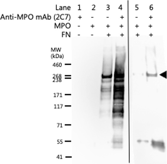
Immunoprecipitation of fibronectin using an anti-MPO mAb. Evidence for a direct MPO-FN interaction was investigated by immunoprecipitation using an anti-MPO mAb (2C7; 2 μg) bound to Protein A/G magnetic beads (Pierce, IL, USA). MPO (20 nM) was pre-incubated with FN (20 nM) to allow potential MPO-FN complex formation prior to incubation with anti-MPO mAb bound to magnetic beads. MPO-FN protein complexes immunoprecipitated with the anti-MPO mAb magnetic beads were separated on 3–8% tris-acetate SDS-PAGE gels and subsequently immunoblotted for the presence of FN monomers (filled arrow) using an anti-FN pAb and imaged by chemiluminescence. Lanes 1–4 were obtained from a blot with a long chemiluminescence accumulation time, carried out to confirm the absence of any recognized material in the controls represented by lanes 1 and 2. The detection of weak bands in lane 3 when compared to lane 4 indicates some non-specific binding of the FN-MPO complex to the beads alone. Lanes 5 and 6, are samples from another experiment, which used a shorter chemiluminescence detection time, to show the differences in staining intensity between the samples with and without mAb 2C7 against MPO. Representative images from n = 3 independent experiments are shown.