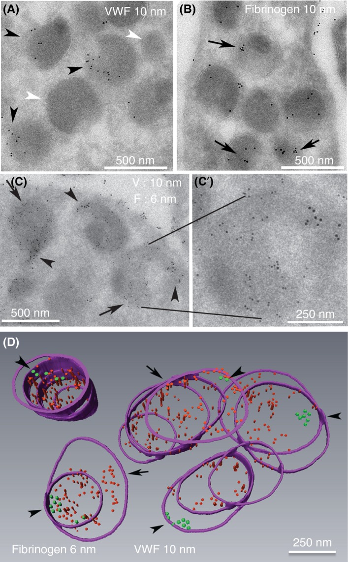
Figure 6.

α‐granules in sequential, serial cryosections immunogold labeled for VWF or fibrinogen exhibited a high labeling incidence. (A, B) Representative 95‐nm cryosections, single immunogold‐labeled for VWF (A, green arrowheads, rabbit polyclonal antibodies, Dako, A0082; **, VWF negative granule profiles) or fibrinogen (red arrows, rabbit polyclonal antibodies, Calbiochem, 341552, MilliporeSigma (Calbiochem), Burlington, MA, USA). The fibrinogen labeling was more common across the granule population; 6‐nm gold particles were used to highlight the fibrinogen labeling shown in C and D, and the larger 10‐nm gold was used for VWF. (C,C’) Double‐label immunogold labeling for VWF and fibrinogen in 95‐nm sections. The double label was done sequentially with a fixation step between the first‐round labeling for fibrinogen and the second‐round labeling for VWF (see Materials and Methods for more details); 2× enlarged single representative α‐granule shown in insert demonstrates abundantly labeled fibrinogen and zoned labeling for VWF. (D) Purple hoops mark the membrane perimeter of the α‐granules as seen in the sequential serial sections. Qualitatively, rendered reconstruction of 5 individual α‐granules immunogold labeled for both fibrinogen (red particles) and VWF (green particles, arrowheads) showed that all were positive for both proteins, with the VWF labeling being restricted to a small zone within each granule. Note the lack of intermixing of fibrinogen and VWF labeling in this sequential, fibrinogen, followed by VWF labeling procedure. With respect to any given granule, the angle of cut may vary, and the full granule volume may or may not fall in the 6‐10 sequential sections of a serial section ribbon. Hence, the number of hoops (perimeter outlines) will vary from granule to granule
