
Figure S10. Quantification of micrometastasis formation in microfluidic devices.
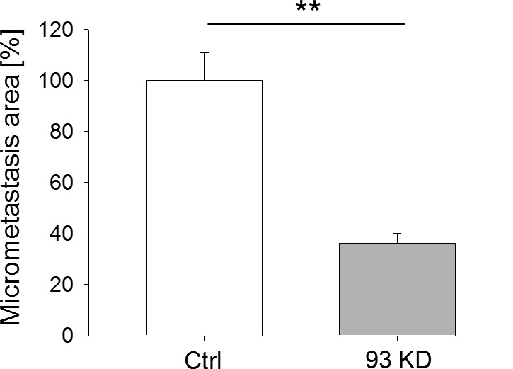
BCCs were seeded in fibrin gel and monitored for 4 d. The ability to form micrometastases was quantified by computing the area covered by fluorescently labeled BCCs at day 4. t test with P < 0.01, N ≥ 10 independent regions in three biological replicates, data normalized to ctrl cells.