
Figure 5.
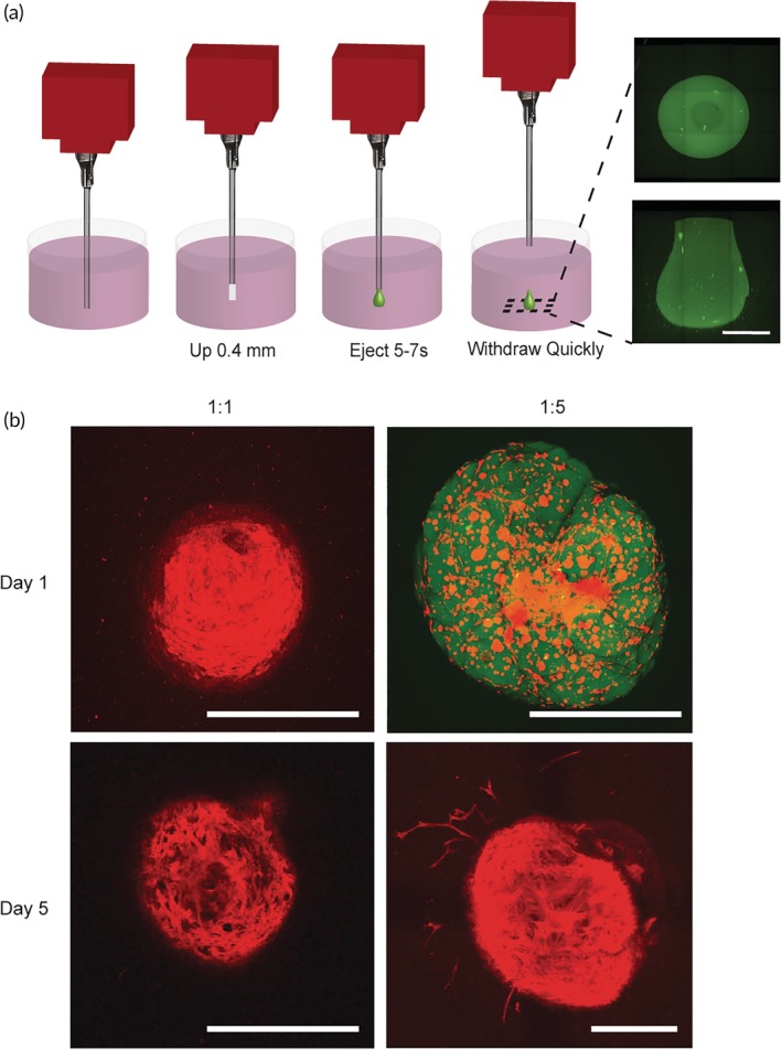
Cell administration studies in vitro. (a) Schematic explaining experimental set‐up of in vitro hydrogel injection experiments. 3D bioprinter is injected in 2.5 mg/ml collagen hydrogel, raised up 0.4 mm, hydrogel is ejected for 5–7 s from the nozzle, and removed quickly out of the collagen hydrogel. Images shown on the right are of a FITC‐labeled 1:5 PNP hydrogel from below (top image) and a sideview (bottom image). Scale bar represents 500 μm. (b) Maximum intensity confocal images of hMSCs encapsulated and delivered in 1:1 and 1:5 PNP hydrogels into collagen hydrogel across a 500 μm z‐stack. Cellular actin is stained with TRITC phalloidin (red) and the HPMC‐C12 is modified with 1 wt.% FITC (green). Images are from below. All scale bars represent 500 μm