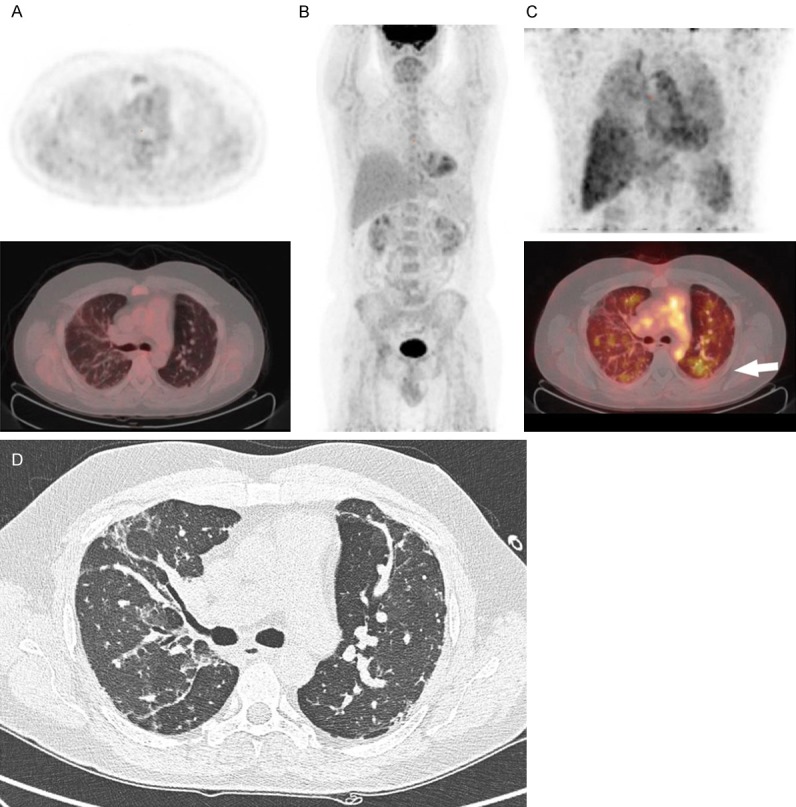
Figure 3.

[18F]FDG PET/CT and [89Zr]Zr-rituximab PET/CT of patient 4. (A) [18F]FDG PET axial views (top: PET, bottom: fusion PET/CT); (B) Maximum intensity projection of [18F]FDG PET; (C) [89Zr]Zr-rituximab PET (top: MIP image, bottom: fusion PET/CT); (D) HRCT or respective axial level of (A and C). 44-year-old patient with antisynthetase associated usual interstitial pneumonia. In the axial views of the [18F]FDG PET image (1 week before rituximab) no increased uptake is seen in the pulmonary regions (perhaps some asymmetry in right versus left lung due to poor aeration) nor in the mediastinal or hilar lymph nodes. The [89Zr]Zr-rituximab PET (C) demonstrated mainly visual diffusely increased pulmonary activity, in contrast to [18F]FDG PET (A, B). The parenchyma shows a moderately increased activity in patchy areas such as in the left lower lobe (white arrow) without any clear anatomical substrate on CT (D). There is normal splenic activity.
