
Figure 1.
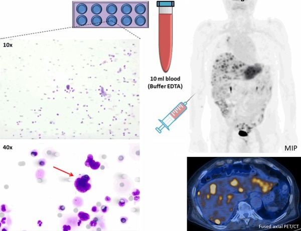
Representative CTC isolated with ISET technique from 10 ml peripheral blood (buffered in EDTA) obtained from a relapsed NSCLC patient with multiple liver metastases. The identified CTC is visualized by MGG staining in 40× magnification (red arrow), and conformed to malignant properties: increased nucleus/cytoplasm ratio, nucleus larger than 3 calibrated pore size of the membrane (>24 μm), irregular nuclear borders, and nuclear hyperchromatism. Round grey spots are 8 μm membrane pores. CTC = circulating tumor cell; MGG = May-Grünwald-Giemsa; ISET = Isolation by Size of Tumor cells; EDTA = Ethylenediaminetetraacetic Acid; MIP = maximal intensity projection.