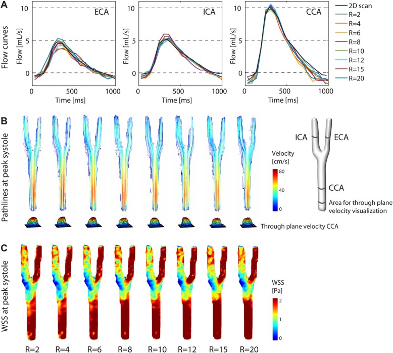
Fig. 5.
Flow in the carotid phantom at (a) external carotid artery (ECA), internal carotid artery (ICA) and common carotid artery (CCA) for acceleration factors R = 2–20 and the fully sampled 2D flow CMR scans. b Pathlines and wall shear stress (WSS) (c) in the carotid phantom for acceleration factors R = 2–20 during peak systole

