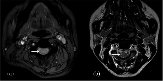
Figure 3.
The spinal cord lesions of NMOSD-associated SSM lesion (a) and MS-associated SSM lesion (b) on T2-weighted imaging. NMOSD-associated SSM lesion was more likely to be centrally located, grey matter involving and transversally extensive on axial imaging.
MS, multiple sclerosis; NMOSD, neuromyelitis optica spectrum disorder; SSM, short segment myelitis.

