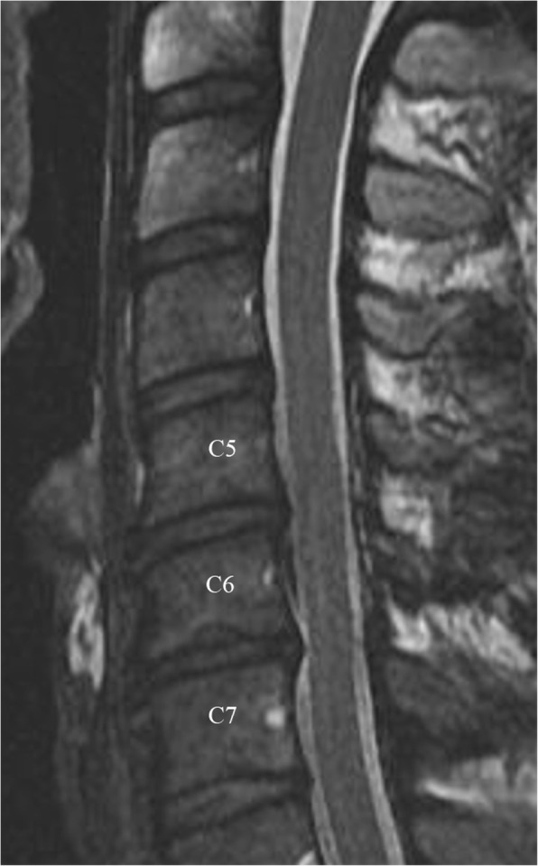
Fig. 3.

Sagittal T2 weighted images of the cervical spine of a male basketball player showing disc dissecation at C5–6 and C6–7. There is also decreased disc space with loss of the differentiation between the annulus fibrosis and nucleus pulposus at C6–7, consistent with Pfirrmann grade IV disc degeneration. Note is made of disc-osteophyte complex at C5–6 and C6–7 and lower C6 Shmorl’s node
