
Figure 2.
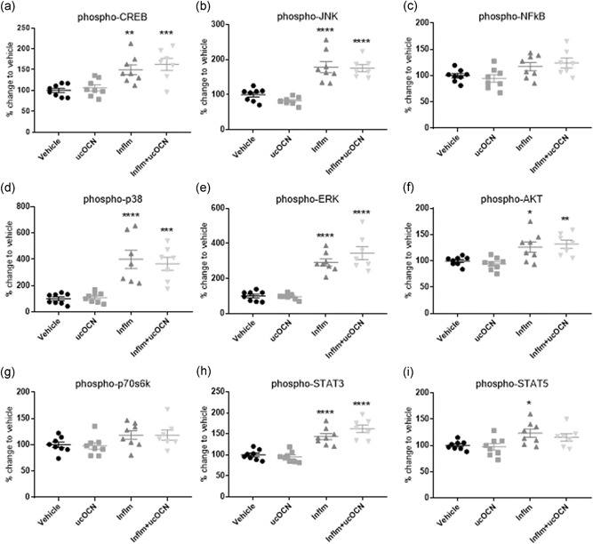
Intracellular signaling responses in HAECs. Luminex® xMAP® technology was used to detect changes in phosphorylated CREB (pS133), JNK (pT183/pY185), NFkB (pS536), p38 (pT180/pY182), ERK (pT185/pY187), Akt (pS473), p70 S6K (pT412), STAT3 (pS727), and STAT5A/B (pY694/699; 48‐680MAG; Milliplex™; Merck Millipore) in cell lysates when treated with vehicle or ucOCN (10 ng/ml) with and without inflammatory stimulus (IFN‐γ and TNF‐α) for 24 hr. Data were analyzed by one‐way ANOVA with multiple comparisons to vehicle corrected for by Dunnett's test. Data are given as means with error bars representing SEM. *denotes a significant difference compared with vehicle (*p < .05. **p < .01, ***P < .005, ****p < .001). HAEC, human aortic endothelial cell; IFN‐γ, interferon‐γ; inflm, inflammatory protocol (8 hr of IFN‐γ 10 ng/ml followed by addition of TNF‐α 10 ng/mL for 16 hr); SEM, standard error of mean; TNF‐α, tumor necrosis factor‐α; ucOCN, uncarboxylated osteocalcin