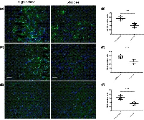
Figure 5.

Impact of l‐fucose on immune cell infiltration after 30 min of ischemia. Individual mice were dosed intraperitoneally with 100 mg of l‐fucose or d‐galactose 1 h before induction of 30 min bilateral renal ischemia and again at the time the clamps were removed. Renal function (BUN) was measured in serum 24 h post‐reperfusion. A, Representative images of immunochemical staining of neutrophils in kidneys sections from mice with l‐fucose or d‐galactose IP injections. B, Quantification of neutrophils in kidney sections from the mice represented in A. C, Representative images of immunochemical staining of leukocytes in kidney sections from mice with l‐fucose or d‐galactose IP injections. D, Quantification of leukocytes in kidney sections from the mice represented in C. E, Representative images of immunochemical staining of macrophages in kidneys sections from mice with l‐fucose or d‐galactose IP injections. F, Quantification of macrophages in kidney sections from the mice represented in E. Scale bars = 100 µm. All images are representative of staining (n = 7 and n = 5 mice/group for d‐galactose and l‐fucose groups respectively). Error bars on all graphs are standard error of the mean. ***P < .005
