Figure 6.

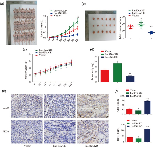
TCONS_00020456 inhibits glioblastoma tumor formation in vivo. (a) U87 cells (2 × 10⁶ cells per mouse; n = 6) were inoculated in BALB/c nude mice to establish subcutaneous xenograft tumors. The mice were euthanized at 41 days and the tumor sizes were measured (*p< .01). (b and d) Photographs of dissected tumors. The relative tumor volume and the tumor weights were measured at 41 days (*p < .01, **p < .01). (c) Mice weight was continuously monitored. (e) Representative images of immunohistochemical staining for smad2, PKCα in different groups. (f) IOD comparison of smad2/PKCα immunohistochemically staining (compared with the vector group, *p < .01, **p < .01). IOD, integral optical density; PKCα, protein kinase C α
