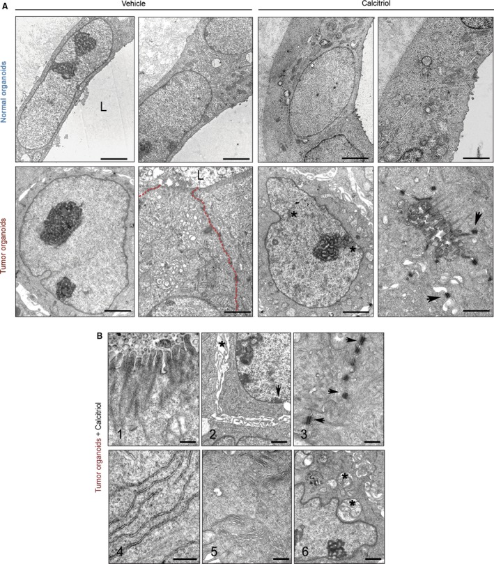
Figure 3.

Calcitriol induces cell differentiation in human tumor organoids. (A) Representative ultrastructural images of normal (patient #11) and tumor organoids (patient #38) treated with 100 nm calcitriol or vehicle for 96 h. Upper panel scale bars (from left to right): 2 μm, 2 μm, 2 μm, and 1 μm. Lower panel scale bars: 2 μm, 1 μm, 2 μm, and 1 μm. L, lumen; asterisks, heterochromatin aggregates; arrowheads, desmosomes; red‐dotted line, intercellular region lacking mature adhesion structures. (B) Epithelial differentiation features induced by calcitriol in tumor organoids from patients #4 and #29. (1) Microvilli. (2) Heterochromatin clumps (arrows) and dilated intercellular spaces (asterisks). (3) Desmosomes (arrows). (4) Rough endoplasmic reticulum. (5) Golgi complexes. (6) Autophagic vacuoles (asterisk). Scale bars (from 1 to 6): 0.4 μm, 1 μm, 0.5 μm, 0.5 μm, 0.5 μm and 1 μm.
