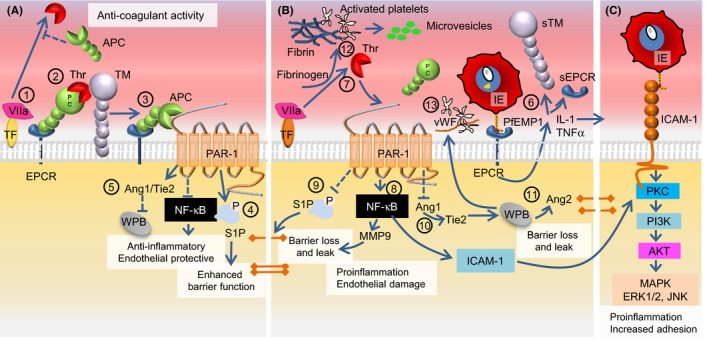
Figure 4.

Linking the protein C pathway with EPCR‐ and ICAM‐1‐binding IEs in cerebral malaria. (A), Effects of EPCR in the absence of Plasmodium falciparum‐IE. Thrombin (Thr) is produced by the interaction between tissue factor (TF) and circulating activated factor VII (VIIa) (1). Thrombin initiates the EPCR‐ and thrombomodulin (TM)‐facilitated activation of protein C (APC) that then inhibits thrombin production (2). APC uses EPCR as a coreceptor for cleavage of proteinase‐activated receptor 1 (PAR‐1). The EPCR‐APC activation of PAR‐1 inhibits the nuclear factor‐κB pathway and exerts anti‐inflammatory and anti‐apoptotic activity (3). S1P signaling results in decreased endothelial permeability, and S1P production leads to enhancement of tight junctions and protection of endothelial barrier integrity (4). Angiopoetin‐1 (Ang1) produced in response to the APC‐PAR‐1 interaction decreases Weibel‐Palade body (WPB) exocytosis by occupying Tie2 (5). (B), The impact of infected erythrocytes expressing EPCR‐bound PfEMP1 on the surface. The IE‐EPCR interaction activates endothelial cells to release pro‐inflammatory cytokines (IL‐1, TNFα) that induce shedding of EPCR and TM from the endothelial surface and increases expression of ICAM‐1 (6). The EPCR‐IE interaction results in reduced levels of APC and increased thrombin generation with fibrin deposition (7). Increased levels of thrombin shift the PAR‐1 response toward activation of the RhoA and NFκB with increased surface expression of ICAM‐1 on the endothelial cell (8). The shift in the PAR‐1 response inhibits S1P release resulting in loss of tight junctions, and compromises endothelial barrier function by causing localized vascular leaks (9). A reduction in Ang‐1 levels increases WPB exocytosis via Tie2 and production of von Willebrand Factor (vWF) and Ang2 (10). Increased levels of Ang‐2 further increase WPB exocytosis and contribute to the loss of endothelial barrier integrity and leakage (11). Platelets become activated by thrombin and cytokines, which leads to production of platelet microvesicles (12). Thrombin and activated platelets combine to form thrombi (13). Strings of vWF and activated platelets form complexes, which like thrombi impair the cerebral circulation. (C), The increase in ICAM‐1 (in panel B) allows IE expressing PfEMP1 with a shared DBLβ ICAM‐1 motif to adhere to the brain endothelium. A large proportion of the ICAM‐1‐adhering IEs might initially bind EPCR via their CIDRα1 domains
