Figure 5.

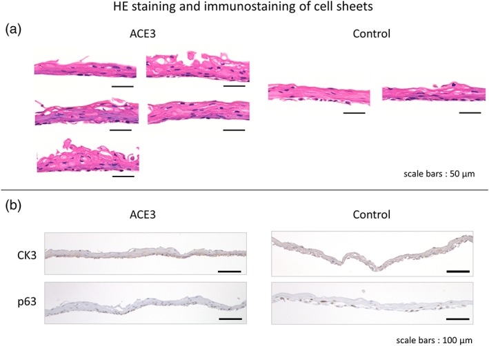
HE staining and immunostaining of cell sheets. HE staining was performed on (a) five samples per test of the automated cell culture, and (b) two samples per test of the manual culture control. The figure of HE staining shows the results of the fourth automated cell culture test. The images of immunostaining were obtained from cell sheets embedded in paraffin blocks using anti‐cytokeratin 3 antibodies and anti‐p63 mouse monoclonal antibodies. The figure of immunostaining shows the results of the fifth automated cell culture test. HE, hematoxylin and eosin [Colour figure can be viewed at http://wileyonlinelibrary.com]
