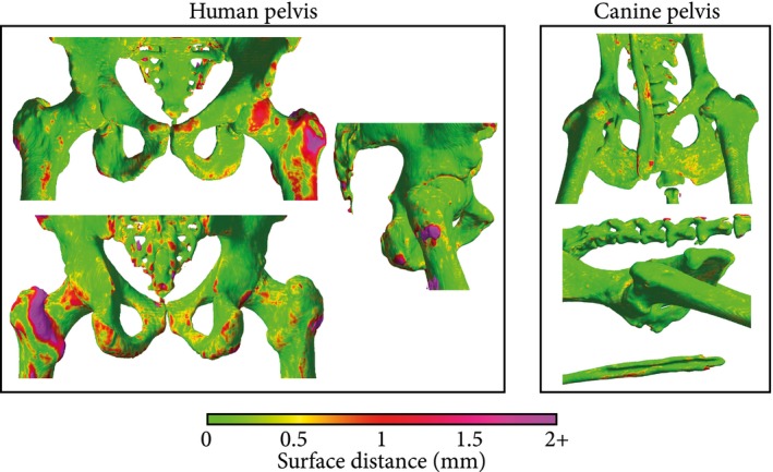
Figure 7.

Surface distance maps obtained for human (subject 8) (A) and canine (subject 13) (B) subjects. Meshes were obtained by thresholding the CT at 200 HU and removing unconnected components. The color map indicates the bilateral surface distance between the CT and a sCT obtained using a Dixon input configuration. The high errors in the human subject, especially in the left femur, originate from registration errors between the CT and the MR/sCT. The canine bone rendering shows the baculum, a thin penile bone present only in a small fraction of the data set that resulted in higher errors for male subjects
