Figure 1.
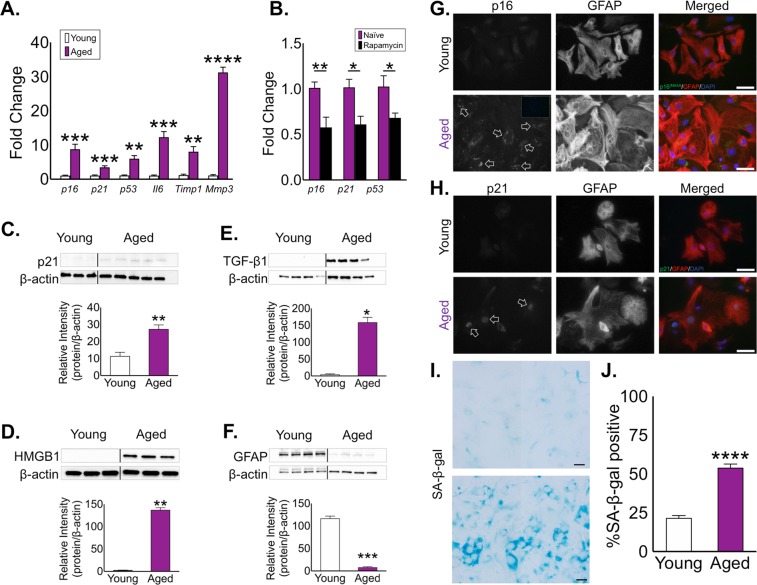
Astrocytes aged in vitro develop a senescence-like phenotype. (A) Analysis of mRNA expression for the senescence-associated genes p16INK4A, p21, p53, Il-6, Timp-1, and Mmp-3 by qPCR in young (white) and aged (purple) astrocyte cultures. (B) Expression of senescence-related genes p16INK4A, p21, and p53 following rapamycin treatment (25 nM/day, 72 h) Fold expression determined by normalization to expression in young astrocytes. Western blot analyses of (C) p21, (D) HMGB1, (E) TGFB1 and the intermediate filament protein (F) GFAP from young and aged astrocyte cell lysates. Densitometry (a.u.) for each factor was used to determine expression relative to β-actin. Representative immunocytochemistry for (G) p16INK4A and (H) p21 in young and aged astrocytes. Scale bar, 20 μm. (I) Representative SA-β-gal staining of young and aged astrocyte cultures, and (J) quantification of SA-β-gal staining in quadruplicate independent cultures. Scale bar, 20 μm. (I) Significance as indicated where: (A) **P < 0.01, ***P < 0.001, and ****P < 0.0001, Student’s t-test with Welch’s correction. (B) *P < 0.05, **P < 0.01, Student’s t-test with Welch’s correction. (C–F) *P < 0.05, **P < 0.01, and ***P < 0.001, Student’s t-test with Welch’s correction. (J) ****P < 0.0001, Student’s t-test with Welch’s correction. Values are expressed as mean ± SEM.

