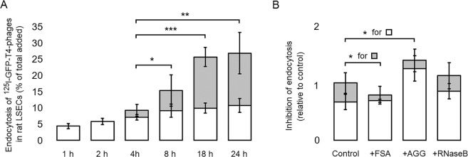
Figure 4.
Endocytosis of T4-phages in LSECs in vitro. (A) Time course endocytosis of 125I-GFP-T4-phages by LSECs. LSEC cultures were incubated with radiolabelled phages for various time periods. For each time point, 3 separate wells containing cells, and 1 cell-free well were used. After each time period, the supernatant from the cells and cell-free well was collected along with one 0.5 ml washing volume of PBS. Trichloroacetic acid (TCA) precipitation was then used to differentiate between free iodine = degraded phages (TCA soluble) (grey columns), and unbound, intact phages (TCA precipitable). Cell bound and internalized phages were quantified in the cell lysates, after solubilizing the cells in 1% SDS (white columns). The results were normalized by subtracting the amount of radioactivity corresponding to the non-specific binding and free 125I in cell-free wells. Each experiment was performed in triplicates, on cells isolated from four animals (Total N = 12 cell cultures for each time point). Bars represent mean ± SD. *p < 0.05, **p < 0.01, ***p < 0.001 represent the statistical differences between the total endocytosis at 18 h and 24 h as compared to 4 h. (B) The specificity of uptake was studied by incubating the cells with 125I-GFP-T4-phages in the absence (Control) or presence of blocking concentrations (0.1 mg/ml) of FSA, ribonuclease B (RNaseB) and aggregated gamma globulin (AGG), inhibitors for stabilin1/2, mannose receptor and FcγRIIb2, respectively. Cell association and degradation were assessed as above. The results are presented as relative uptake compared to the control which was set to 1. Each experiment was performed in triplicates, on cells isolated from 3 animals (Total N = 9 cell cultures for each time point). Bars represent mean ± SD.

