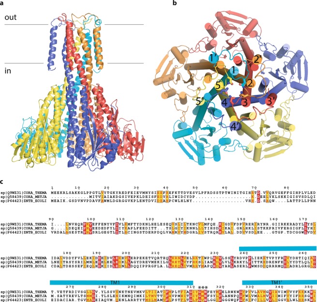
Figure 1.
The general organization of CorA family of proteins as exemplified by TmCorA (pdb code 4I0U). (a) side view, each protomer is color-coded; the position of membrane is indicated with black lines; (b) view from the extracellular part, transmembrane helices are numbered with the numerals, those with ′ indicate the outer transmembrane helix of each protomer. The asparagine side chains of GxN motif are shown as sticks; (c) the sequence alignment of TmCorA, MjCorA and EcZntB. Essentially conserved amino acids are in red. The turquoise bars show the position of the long helices (numbered in panel b) forming the channel and of the periphery helices (indicated with ′ in panel b). The signature motif/selectivity filter is indicated with *. The sequence alignment was produced with T-coffee42 (http://tcoffee.crg.cat/apps/tcoffee/index.html) and annotated with Espript 3.0 (ref. 43) (http://espript.ibcp.fr).

