Figure 2.
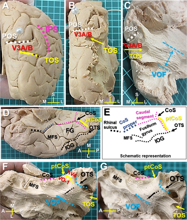
VOF’s cortical projections in the dorsal and ventral visual cortex (Sample #1). (A) Caudal view of the right hemisphere. TOS is located at the bottom of IPS inferior to POS. (B) Caudal view after removal of the cortex to expose the area around TOS. (C) Further dissection to expose the VOF’s cortical projections into TOS. (D) Ventral view of the right hemisphere with representative anatomical landmarks. (E) Schematic representation of ventral temporo-occipital cortex to show the CoS complex (Rhinal sulcus, CoS proper, and caudal segment), ptCoS, OTS, IOG, and fusiform gyrus (FG). (F) Ventral temporal lobe after removal of the cortex to expose the white matter around OTS and ptCoS. (G) Further dissection to expose the ventral VOF’s cortical projections in posterior fusiform gyrus and ptCoS. IPS; intraparietal sulcus, POS; parieto-occipital sulcus. CC; corpus callosum, CoS; collateral sulcus, ptCoS; posterior transverse CoS, FG; fusiform gyrus, MFS; mid-fusiform sulcus, IOG; inferior occipital gyrus, OTS; occipito-temporal sulcus, TOS; transverse occipital sulcus. A; anterior, L; lateral, M; medial, P; posterior, S; superior.

