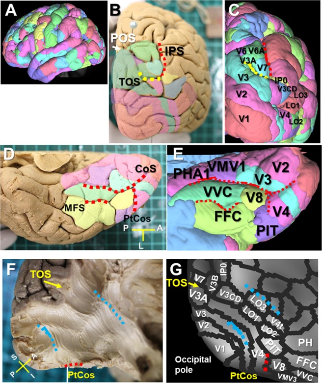
Figure 3.
VOF’s cortical projections on the HCP MMP1.0 atlas. (A) Lateral view of HCP-1021 template overlaid with HCP MMP1.0 atlas. (B,C) Caudal view of a brain sample (B) and HCP-1021 template (C) with HCP MMP1.0 atlas. (D,E) Ventral temporal cortex of brain sample (D) and HCP-1021 template (E) with HCP MMP1.0 atlas. (F,G) Posterolateral corner of a brain sample (F) after dissection to expose the VOF’s fiber bundles running between ventral and dorsal visual stream, with corresponding HCP MMP1.0 atlas in a surface-based coordinate system (G). IPS; intraparietal sulcus, TOS; transverse occipital sulcus, POS; parieto-occipital sulcus. CoS; collateral sulcus, ptCoS; posterior transverse CoS, FG; fusiform gyrus, MFS; mid-fusiform sulcus, OTS; occipito-temporal sulcus. A; anterior, I; inferior, L; lateral, P; posterior, S; superior.

