
Figure 7.
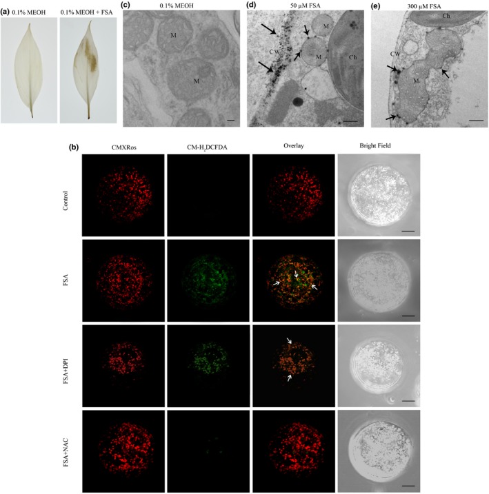
Reactive oxygen species (ROS) detected in fusaric acid (FSA)‐treated banana leaves and embryogenic cell suspension (ECS) protoplasts. (a) Leaves were treated with 0.1% MEOH or 300 μM FSA and stained with DAB as described in the Materials and Methods section. The brown precipitate indicated DAB polymerization at the site of ROS production. (b) Confocal laser scanning microscopy of protoplasts from 7‐d‐old banana ECSs treated with 0.1% MEOH (control), FSA (300 μM), FSA + DPI (inhibitors of NADPH oxidase), and FSA + NAC (a ROS scavenger) for 5 min. Samples were double‐stained with CM‐H2 DCFDA (to detect ROS) and CMXRos (mitochondrial marker). (c) to (e) Electron microscopy localization of H2O2 production in banana tissue culture seedlings treated with 0.1% MEOH (c), 30 μM FSA (d) or 50 μM FSA (e). Three or more different leaf samples of both 0.1% MEOH‐treated and FSA‐treated plants were used in each experiment. Images represent typical observations in two independent experiments. Arrows indicate depositions of cerium. Bars: (b) 10 μm; (c) 200 nm; (d, e), 1 μm. M, mitochondrion; CW, cell wall; Ch, chloroplast.