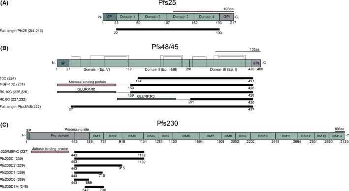
Figure 4.

Native protein structure of Pfs25 (A), Pfs48/45 (B) and Pfs230 (C). (A) Schematic representation of the four EGF‐like domains of Pfs25 with 22 cysteines with, underneath, the full‐length vaccine construct used in preclinical and clinical studies. (B) Domain structure of Pfs48/45 with cysteines forming disulphide bridges (dotted lines) based on homology to other 6‐cys domain proteins. Underneath, several vaccine constructs are presented that have been tested in preclinical studies. (C) Schematic of Pfs230 with 14 cysteine motifs (CM). The processing site is the location where the protein is cleaved after gamete emergence from the red blood cell. Underneath, vaccine constructs that have been tested in preclinical studies; Pfs230D1M has been tested in clinical studies (ClinicalTrial.gov NCT02334462 and ClinicalTrial.gov NCT02942277). SP: Signal peptide; GPI: Glycosylphosphatidylinositol anchor
