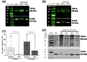
Figure 2.

Expression levels of TRPV4. A, GP tissue; Representative western blot of TRPV4 (98 kDa), Abcam primary antibody. B, GP tissue; Representative blot of TRPV4 (98 kDa), Alomone primary antibody. β‐actin loading controls (42 kDa) shown in blot below dotted blue line. (RBL: rat brain lysate Muc: mucosa, SM: smooth muscle). Membrane was cut at 55 kDa (dashed blue line) and each half probed with the appropriate antibodies. C, Quantified data of TRPV4 expression by densitometry, normalized to its loading control. Samples duplicated and averaged. Results for each tissue type are similar when using either primary antibody; mucosal lysates have significantly higher TRPV4 expression than in smooth muscle lysates with either primary antibody. No significant difference between antibodies for either tissue type. Median values [25, 75], Abcam; n = 5, Alomone; n = 8, Mann‐Whitney. D, Western blot of human mucosal biopsies. Three of the four samples assessed have a positive band at 98 kDa confirming presence of TRPV4. One sample is negative for TRPV4; however, this is likely due to sample degradation due to reuse
