
Figure 3.
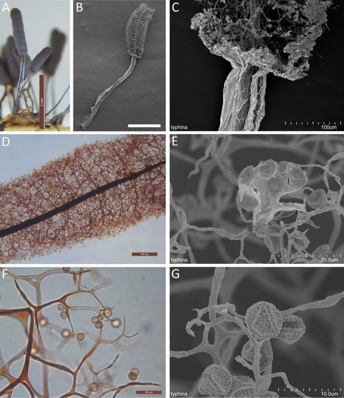
Stemonitopsis typhina. A. Sporocarps grew on an agar medium. B. Sporocarps by SEM. C. The base of sporotheca shows the continuous of peridium with the membrane surrounding its stipe. D. Part of sporotheca by transmitted light. E. Peridial fragment in the surface of sporotheca covers several spores by SEM. F. Spores by transmitted light. G. Spore by SEM. Scale bars: A = 2 mm; B = 1 mm; C, D = 100 μm; E, F = 20 μm; G = 10 μm.