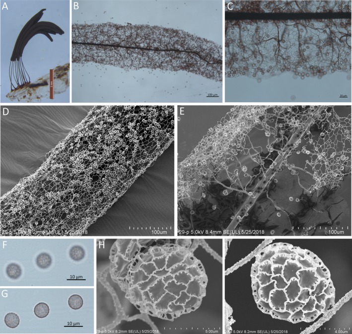
Figure 6.

Stemonitis fusca. A. Sporocarps grew on an agar medium. B. Part of sporotheca by transmitted light. C. Part of the surface net by transmitted light. D. Part of sporotheca shows surface net by SEM. E. Part of sporotheca shows capillitium by SEM. F, G. Spores by transmitted light. H, I. Spore by SEM. Scale bars: A = 2 mm; B, D, E = 100 μm; C = 20 μm; F, G = 10 μm; H = 5 μm; I = 4 μm.
