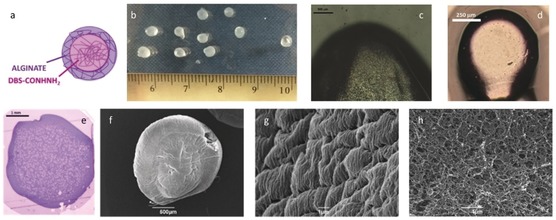
Figure 3.

Images of hybrid DBS‐CONHNH2/alginate gel beads. a) Schematic diagram of a bead; b) Photograph of beads (droplet size 20 μL) adjacent to a ruler (scale in cm); c)–e) Optical microscopy of the cross‐section of a bead. c) Droplet size 20 μL, scale bar 500 μm, d) droplet size 1 μL, scale bar 150 μm, e) Bead embedded in resin and coloured using toluidine blue (scale bar 1 mm); f) SEM image of a bead (scale bar 500 μm); g) SEM image of the surface of a bead (scale bar 1 μm); h) SEM image of the interior cross‐section of a bead (scale bar 1 μm).
