
Figure 2.
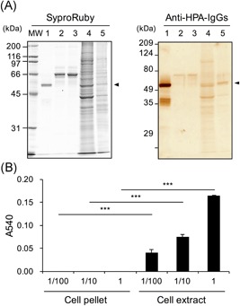
Western‐blotting for α‐amylase and starch‐degrading activity in Caco‐2 cells and culture medium. Caco‐2 cells were seeded at 5 × 10⁴ cells/cm2 in dishes (150 × 20 mm) and cultured for 6 days. The cells and culture medium before and after culture (2 mL each) were prepared as described in Section 2. A, Western‐blotting for α‐amylase: The samples were boiled with SDS‐PAGE sample buffer including 2‐mercaptoethanol, and 10 μL aliquots were used for Western blot analysis as described in Section 2. Left, Protein staining using Sypro Ruby. Right, Immunostaining for α‐amylase using rabbit anti‐HPA IgGs and HRP‐conjugated goat anti‐rabbit IgGs, MW: molecular weight maker, Lane 1, purified pig pancreatic α‐amylase (0.8 μg/lane); 2, medium before culture (1.7 μg protein/lane); 3, medium after culture (1.8 μg protein/lane); 4, cell pellet (12 μg protein/lane); 5, cell extract (2.3 μg protein/lane). B, Starch‐degrading activity: Caco‐2 cells were cultured in dishes (150 × 20 mm) for 6 days. The cell pellet and extract were separately solubilized in 500 μL of 20 mM phosphate buffer, pH 6.9. The protein concentrations of the cell pellet and extract were 0.227 and 1.16 mg/mL, respectively. The cell pellet and extract (1, 10, and 100 times dilution) were used as enzyme samples (1 vol was 15 μL) as described in Section 2. HPA, human pancreatic α‐amylase; IgG, immunoglobulin G; SDS‐PAGE, sodium dodecyl sulfate‐polyacrylamide gel electrophoresis ***P < 0.001 vs cell pellet by unpaired t test